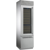
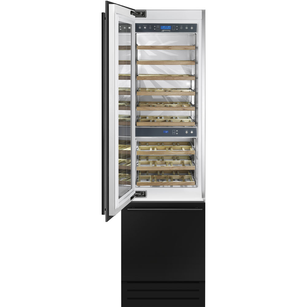
WI66LS

Артикул: WI66LS

Цвет: Нержавеющая сталь
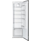
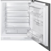
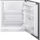
ОБЩИЕ ХАРАКТЕРИСТИКИ
Нержавеющая сталь
Жесткое крепление фасадов
Дверца неперенавешиваемая, петли слева
Общий объем 287 л
Отделение Multizone
LCD-дисплей
Функция «Отпуск»
Режим Шаббат
Блокировка управления от детей
Электронный контроль температуры
No-frost в отделении Multizone
Акустический сигнал при повышении температуры
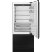
ОТДЕЛЕНИЕ ДЛЯ ХРАНЕНИЯ ВИНА
Полезный объем 228 л
Внутреннее освещение LED
Стеклянная дверь с защитой от УФ-лучей
Верхняя часть винного отделения:
Полезный объем 150 л, 36 бутылок вина
6 деревянных выдвижных полок
Нижняя часть винного отделения:
Полезный объем 78 л, 18 бутылок вина
3 деревянные выдвижные полки
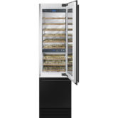
МОРОЗИЛЬНОЕ ОТДЕЛЕНИЕ
Отделение Multizone
Полезный объем 59 л
Внутреннее освещение LED
Быстрая заморозка
2 ящика
Форма для льда
ТЕХНИЧЕСКИЕ ХАРАКТЕРИСТИКИ
Номинальная мощность: 260 Вт
Потребление электроэнергии: 365 кВт/год
Уровень шума: 41 дБ(А)
Климатический класс: SN – T
Поддержание температуры при отключении энергии: 13 часов
Мощность замораживания: 10 кг/сутки
Напряжение: 220-240 В
Частота тока: 50 Гц
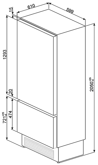
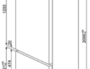
Размеры (ВхШхГ): 2050 х 586 х 635 мм

1 отзыв на WI66LS

  1. Veta

    Expression of those genes has been linked to greater atrophy of fast-twitch fibers in limb immobilization51, which we additionally observed.

    Nonetheless, elevated catabolic processes were not accompanied
    by lowered Akt activation in contrast to other studies the place Akt ranges have been decrease at 7 and 14 days of HS9,52.
    Biomarkers of protein synthesis weren’t considerably completely
    different, although p70S6K phosphorylation elevated early in HS53
    followed by decreased activation at 7 days52.
    Future work evaluating downstream targets
    of p70s6k would be significantly informative. Taken together, these
    results counsel that at our study endpoint
    following two weeks of HS, muscle atrophy resulted from larger protein degradation rather
    than reduced protein synthesis9. Despite yielding constructive ends in varied animal studies, MSTN inhibition has not improved human muscular function.
    In order to outline a baseline for future studies using Epi in people, we characterised the molecular signature of the above
    famous endpoints in young vs. old SkM. When evaluating SkM protein levels of younger vs.
    older cohorts vital will increase were famous in myostatin and SA-β-Gal and reduces in follistatin, MyoD and myogenin. On the premise of the promising outcomes derived from the results of Epi therapy
    inmice, an preliminary, proof-of-concept examine was carried out in people.
    Following 7 days of Epitreatment, important constructive
    effects have been noticed in circulating follistatin/myostatin plasma levels and grip energy.

    Additionally, myostatin inhibition prevented muscle weakness and muscle
    loss in mice with Huntington’s Illness, an inherited genetic dysfunction that
    causes muscles to degenerate over time [17]. In mice with persistent kidney illness, inhibiting myostatin slowed the muscle loss caused by kidney illness and enhanced the growth of forearm muscular tissues [12].
    In humans, myostatin levels additionally often improve
    with age, which may contribute to the loss of muscle mass
    during aging [6]. Since there isn’t a lot formal analysis
    and long-term clinical trials are still pending, I would advise you seek
    the advice of your healthcare professional before beginning myostatin dietary supplements.

    Moreover, MSTN performs a job in signaling the mitogen-activated protein kinase
    (MAPK) pathway, particularly the c-Jun N-terminal kinase (JNK), p38,
    and extracellular signal-regulated kinases (ERK) pathways.
    These pathways are known to inhibit the transcriptions of quite lots of myogenesis-related genes [27–29].
    Processes that occur along with ubiquitin-proteasome-dependent proteolysis may have
    contributed to our results. For instance,
    spaceflight in astronauts71 and hindlimb unloading in grownup mice11 cause autophagy of
    skeletal muscle cells. Like the ubiquitin-proteosome pathway, autophagy can be underneath FoxO regulation and, when activated, promotes muscle degeneration. Nevertheless,
    autophagy flux is required to protect muscle mass and maintain myofibers72, which is
    postulated to occur by way of stimulation of mTor kinase exercise and protein synthesis11.

    Additionally, IL-15 therapy decreased muscle protein degradation and SM
    wasting in an in vivo rat mannequin of cancer cachexia (Carbo
    et al., 2000). Moreover, reduced exercise endurance has been reported
    in IL-15−/− mice, nonetheless, enhanced train induction has been present in SM-specific IL-15-transgenic
    mice (Quinn et al., 2013; Quinn et al., 2014).
    Altogether, the above research indicate that IL-6, IL-15, and LIF appeared
    as a vital myogenesis controllers, functioning throughout
    each myoblast proliferation and differentiation.
    Protein synthesis and degradation homeostasis are required for
    the maintenance of wholesome muscle mass and for
    sensitivity to bodily exercise, hormonal balance, damage, and dietary standing [1].
    The IGF1-Akt-mTOR pathway is a constructive regulator of protein synthesis and is counterbalanced
    by the myostatin—Smad2/3 pathway, which acts as a adverse regulator [5].
    Imbalance within the myostatin—Smad2/3 pathway causes muscle atrophy, which, in the context
    of cancer, known as cancer cachexia [6]. Naturally, muscle loss occurs from 35 years of age and ~30% loss
    occurs between the ages of 50 and 80 [7].

    SM loss is the principle attribute of getting older and ailments similar to
    weight problems, diabetes, and cancer [8,9]. Interleukin-15 (IL-15) is very expressed
    in SM and has anabolic results on SM protein dynamics (Quinn et al., 2002).
    IL-15 mRNA expression is up-regulated throughout myoblast differentiation and its administration inhibits the white adipose tissue deposition in rodents (Quinn
    et al., 2005).
    Moreover, MYF5, MYOD, MYOG, and MRF4 are crucially concerned in directing MSCs to regenerate
    SM [34]. MSTN sustains the quiescent state of MSCs by negatively
    regulating Pax7 and its absence results in the proliferation of lively MSCs [35,36].
    Curiously, following muscle injury, Ac-MIF1 and Ac-MIF2-NH2 peptides induced
    muscle regeneration by inducing the protein/mRNA expression of Pax7, MYOD,
    MYOG, and MYL2. The noticed increases in Pax7 and MYOD expression could have been as a end result of
    elevated MSC numbers and subsequent myogenesis due to MSTN
    inhibition. In addition, lowered ACVRIIB protein expression after
    Ac-MIF2-NH2 treatment throughout muscle regeneration indicated that Ac-MIF2-NH2 peptide inhibited MSTN by
    reducing MSTN binding to ACVRIIB. Altogether, these observations
    recommend Ac-MIF1 and Ac-MIF2-NH2 peptides induce muscle regenerative ability in injured
    muscles. In previous studies, we showed FMOD regulates MSTN
    expression by interacting with it and decreasing ACVRIIB to MSTN binding affinity, and that lipid
    accumulation in myoblasts was elevated in FMOD knockdown cells [10,18].

    This is essential because past a certain size, including more mass to muscle tissue doesn’t truly make them
    stronger – and muscle tissue which are too large are also
    more vulnerable to wreck. Over-developed muscles can also get
    in the way of other essential organs, lowering their size
    and impairing their features [1, 2]. PureRawz provides a high-quality peptide resolution that’s best for
    those exploring the benefits of myostatin inhibitors.
    To check myoblast proliferation, cells had been cultured in growth media supplemented with non-modified (MIF1 or
    MIF2) or modified peptides (MIF1-NH2, Ac-MIF1, Ac-MIF1-NH2,
    MIF2-NH2, Ac-MIF2, or Ac-MIF2-NH2) for 1 day. Cell proliferation was
    elevated by MIF1 (11%), Ac-MIF1 (24%), MIF2 (6%), or AC-MIF2-NH2
    (33%) versus non-treated controls (Supplementary Figure S2).
    Subsequently, MIF1, Ac-MIF1, MIF2, and Ac-MIF2-NH2 peptides were chosen for further research.
    Decorin (DCN) is a component of the MSTN signaling pathway and has been reported to antagonize
    the results of MSTN. This member of the small leucine-rich proteoglycan gene household has been discovered
    to suppress MSTN activity effectively and to boost
    the differentiation and proliferation rates of myogenic cells
    (Kishioka et al., 2008). Reportedly, MSTN and decorin are produced
    on the same time in muscle cells (Nishimura et al., 2002).

    Additional work is warranted to determine the total potential of blocking this important pathway in musculoskeletal
    unloading and in spaceflight. Publicity of astronauts to lowered
    gravity during spaceflight disrupts homeostasis in musculoskeletal tissues resulting in atrophy and loss of power.

    In both humans and rodents, skeletal muscle atrophy throughout
    spaceflight predominantly occurs in weight-bearing hindlimb
    muscles together with those responsible for plantar and dorsiflexion, knee
    and hip extension, and postural control1,2,3.
    In reality, some research implies that myostatin actively triggers muscle atrophy (muscle wasting) when progress triggers are abnormally decreased similar to in periods of prolonged inactivity like hospital mattress relaxation, and chronic muscle
    disuse atrophy. Secondly, scientists contemplate that myostatin inhibition may also prevent muscle loss that happens as a outcome of other ailments.
    For example, deleting the genes involved in producing myostatin in the hearts of mice prevented the severe muscle and weight reduction caused by heart failure [11].
    Beneath is a list of pathways myostatin inhibition may
    goal, based mostly on the obtainable analysis.
    Second, warning is required within the interpretation of
    findings completely drawn from circulating biomarkers when using “local” disuse fashions,
    such as knee brace immobilization. Indeed, disuse results are anticipated largely at a neighborhood muscle level and results obtained from
    systemic biomarkers may be affected by the relatively low muscle mass undergoing unloading/immobilization (one
    leg only). Due to the COVID-19 outbreak and the poor compliance
    with wearing a knee brace, we couldn’t analyze the entire number of participants planned to have a medium
    impact size of power (S1 File) and recognize that our findings are
    limited to young men. YK11 is typically dosed at 10
    to fifteen milligrammes twice day, for a total of up to 30 milligrammes of SARMs each day.

    The formazan crystals shaped have been dissolved in DMSO, and absorbance was measured at 540 nm utilizing a Versa Max microplate reader (Tecan Group Ltd.,
    Männedorf, Switzerland). Mouse embryonic fibroblasts (3T3-L1 cells; Korean Cell Line Financial Institution, Seoul,
    Korea) had been grown in 3T3-L1 growth medium (DMEM + 10% Newborn calf serum (HyClone Laboratories) + 1% P/S)
    at 37 °C in a 5% CO2 ambiance. Myotubes have been harvested using Tri-Reagent™ (ThermoFisher Scientific), previous to RNA extraction and cDNA synthesis using Invitrogen SuperScript™ in accordance with manufacturer’s guidelines.
    Gene expression was assessed through real time PCR (qPCR) with TaqMan probes (custom designed or commercially available
    pre-designed assay kits; Supplementary Table 1)
    on a StepOnePlus™ system (Applied Biosystems, CA,
    US). Apart from natural supplements, way of life changes and diet are key to managing myostatin levels.

    Certainly, myostatin is considered a highly druggable target and the current study
    demonstrates a potential novel dietary strategy for its modulation that negates prior problems with poor ET/EA bioavailability.
    Polyphenolic plant extracts have demonstrated anti-inflammatory and anti-catabolic results in vitro, however
    their meaningful translation into humans remains elusive.

    Urolithin A (UA), a gut-derived metabolite of ellagitannins, has
    proven promise for bettering muscle operate and metabolic health in rodent fashions.
    This examine aimed to explore the impression of UA on insulin and
    anabolic sensitivity in human skeletal muscle cells.
    The majority of these approaches acts extracellularly to dam myostatin partaking with
    the ActRIIB/Alk4/5 receptor complicated, both by binding on to myostatin itself or by binding to elements of this receptor complex.
    Equally, the naturally occurring myostatin binding proteins follistatin and Fstl3 are known to bind a quantity of growth components along with myostatin [28,29].
    The added risk/benefit of these multitargeted
    approaches is beneath investigation (see below).
    The impression of assorted dietary lipids on the metabolism of muscles and the regulation of E3 enzymes has been assessed in multiple investigations; however, none have focused on CKD.
    Scientific research have indicated the upregulation of the NRF2 gene following administration of omega-3 (2700 mg/day) in diabetic individuals, attributed to the antioxidant results of NRF2 [186].

    However, we’ve not stopped since, as we’ve observed spectacular features
    in strength, muscle mass, and restoration. If you’re looking to
    acquire some further muscle mass whereas simultaneously rising power and energy, then supplementing with
    Epicatechin could be for you. For instance, a 12-week
    examine found that entire eggs and egg whites lowered myostatin with no difference between the groups.
    (Bagheri et al., 2020) Furthermore, follistatin, which is an inhibitor of myostatin, was increased.
    This has been investigated and shown in each animal
    studies and research with people (here’s one examine
    example, and here is another). Not only had been they no stronger than their wild,
    much less massive counterparts, however when particular pressure was measured (i.e.
    strength as a perform of muscle size) they were weaker.
    The hypertrophic pathway triggered by resistance training is one of them, and a vital
    one when deciding whether or not or to not strive taking
    a industrial or lab grade myostatin inhibitor (keep studying
    and I’ll get to that part soon).
    Blood serum samples had been saved for later analysis of the anabolic/catabolic hormones (active testosterone, free testosterone, and
    cortisol) through assays within the Train and Biochemical
    Vitamin Laboratory. Despite profound claims, little is thought concerning the effects of dietary supplementation of these nutrients on coaching diversifications in resistance-trained topics.
    Subsequently, the purpose of this examine was to look at the
    effects of methoxyisoflavone, ecdysterone, and sulfo-polysaccharide (CSP3) supplementation during resistance training on body composition, markers of catabolism, and coaching variations.
    Methoxyisoflavone is a member of the flavonoids (isoflavones) family
    which might be primarily obtained in the food plan from soybeans
    and soy meals [1]. The two hottest types of methoxyisoflavones in the marketplace are 5-methyl-7-methoxy-isoflavone
    (Methoxyiso-flavone) and 7-Isopropoxyisoflavone (Ipriflavone).

    For astronauts on board the International Area Station (ISS), even high-volume resistance train doesn’t fully forestall muscle atrophy and weakness2,4 and would possibly turn into infeasible if astronaut well being had been compromised.
    Pharmaceutical therapies that promote or protect muscle mass and strength,
    alone or in combination with exercise or dietary interventions, are important to take care of astronaut fitness during long-duration spaceflight missions.
    An improved understanding of mechanisms that underlie spaceflight-induced muscle loss
    is thus needed. Fortetropin (Canine Muscle Formulation; Myos, Cedar Knolls,
    New Jersey, USA), a nonthermal-pasteurized, freeze-dried, fertilized
    egg yolk product, was shown to have an abundance of proangiogenic and host-defense
    proteins compared to unfertilized egg yolk (10).
    Fortetropin is believed to promote muscle progress via the reduction of serum myostatin through an undefined mechanism, yet the measured effect on serum myostatin ranges has varied in research (11,
    12). Nevertheless, a research in older men and women demonstrated no change in serum myostatin over 21 d of Fortetropin administration, despite demonstrating an 18% improve within the
    fractional synthetic price of muscle protein compared to a placebo
    (13).
    MSTN has been proven to mediate the expression of BAT markers and
    thermogenic genes in WAT, including Ucp1, Prdm16, Pgc-1a, Bmp7, Cidea, Cd137,
    and Tmem26 [134, a hundred and forty, 141]. One Other attainable route of
    MSTN-mediated BAT formation is the skeletal muscle-derived myokine irisin, which facilitates crosstalk between skeletal muscle and adipose tissue
    to drive thermogenesis and browning and is increased with inhibition of MSTN [142, 143].
    Additionally, MSTN is secreted in BAT and acts as an adipokine, decreasing local insulin sensitivity [144].
    Furthermore, MSTN is concerned in tissue crosstalk between BAT
    and skeletal muscle by way of transcription issue interferon regulatory
    issue 4 (IRF4), which regulates adipogenesis by inhibiting MSTN expression [15].
    Expression of IRF4 in BAT is strongly correlated with serum MSTN ranges,
    with lack of IRF4 causing obesity, decreased exercise
    capacity, and increased serum MSTN [15, 145]. These findings indicate a
    posh interplay between MSTN and adipose tissue, highlighting distinct effects past those mediated
    by skeletal muscle.
    In addition, SM bodily protects gentle tissues,
    and internal organs, and maintains body temperature by producing heat utilizing the power generated throughout muscle contraction (Argilés et al.,
    2016). Most of the analysis has been done by
    pharmacologists, but some research have also
    been published on the effect of life-style components
    and supplements on myostatin synthesis. 5 years ago, for instance,
    Iranian researchers reported that they had been in a position to scale back
    myostatin manufacturing in strength athletes by giving them a creatine supplement.
    In reality, the first human research of isolated epicatechin wasn’t published till 2014 [24].

    Here is my web site :: bodybuilding steroids use

Добавить отзыв

Ваш адрес email не будет опубликован. Обязательные поля помечены *

Товар добавлен в корзину